The Exposome Scan Facility

Background

Human health is shaped not by single exposures but by the totality of chemical, biological, and lifestyle factors encountered across a lifetime — the exposome. Traditional health studies have approached this complexity one chemical at a time, a strategy that is inherently incomplete and poorly suited to capturing the mixture effects, interactions, and cumulative burden that characterise real-world exposure. The Exposome Scan was established to address this gap: to measure the human chemical environment comprehensively, simultaneously, and at population scale.

About the Facility

The Exposome Scan is a collaborative research infrastructure between Leiden University (Leiden Academic Centre for Drug Research / LACDR) and Utrecht University (Institute for Risk Assessment Sciences / IRAS), designed to enable high-throughput, untargeted measurement of the human exposome in biological matrices. By combining world-class analytical chemistry with epidemiological expertise, the facility bridges the distance between a biological sample and a meaningful health insight.

What It Measures

The facility applies advanced mass spectrometry — primarily high-resolution liquid chromatography–mass spectrometry (LC-HRMS) and gas chromatography–mass spectrometry (GC-MS) — to biological matrices including blood (serum and plasma), urine, and tissue. The goal is to capture as wide a chemical “fingerprint” as possible in a single sample: endogenous metabolites, environmental contaminants (pesticides, persistent organic pollutants, plasticisers, flame retardants, polycyclic aromatic hydrocarbons), pharmaceuticals, and dietary components simultaneously, rather than measuring targeted compounds one by one. Different scans have been developed to target at different depth both exogenuous and endogenuous compounds. Additional, technologies allow for sensitive measurements by for example conctrating with in-line evaporators.

In a single analytical run, the platform can detect and semi-quantify thousands of chemical features across a dynamic concentration range spanning several orders of magnitude. Spectral data are annotated against curated reference libraries and continuously updated as new compounds are characterised, ensuring that the facility’s coverage grows over time. Where full identification is not yet possible, unknown features can be retained and revisited as databases expand — meaning that no signal is discarded prematurely.

Division of Expertise

Leiden brings deep analytical chemistry strength — method development, instrument operation, quality control, and spectral library annotation. The Exposome-Scan facility is hosted by the Dutch Metabolomics Center. Utrecht contributes epidemiological study design, exposure science, and the statistical frameworks needed to link complex multi-exposure profiles to health outcomes in large cohorts. Together this covers the full pipeline: from biobank sample preparation and extraction, through high-throughput mass spectrometric acquisition, to feature detection, annotation, mixture analysis, and biological interpretation. This integrated pipeline reduces the risk of losing signal between analytical and epidemiological stages, which has historically been one of the main bottlenecks in exposome research.

Why It Matters Scientifically

The Exposome Scan approach allows researchers to work in an agnostic, hypothesis-generating mode — identifying which of thousands of detected features associate with a disease outcome — before drilling down into specific compounds for targeted follow-up. This is particularly relevant for diseases where multiple environmental contributors are suspected but no single agent fully explains the observed patterns, such as neurodegenerative diseases, cardiovascular conditions, metabolic disorders, and adverse reproductive and developmental outcomes.

Beyond environmental epidemiology, the facility supports pharmaco-exposomics applications — examining how ph
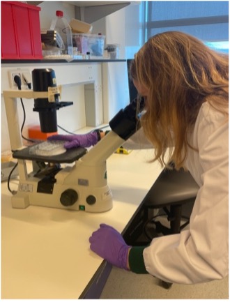
The Princess Máxima Imaging Center plays a crucial role in pediatric cancer research by providing state-of-the-art imaging technology and expertise. Microscopy plays a vital role in visualizing the morphology, behavior, and activity of cells over time, enabling a deeper understanding of spatial and temporal dynamics in complex biological systems. By investing in microscopy, researchers gain a unique perspective on the interactions and processes within 3D organoids. This facilitates the study of human biology, drug screening, and examination of cancer and environmental cell interactions, particularly in the context of immunotherapies.

Expert knowledge and support accelerate research through guidance in experimental design, image processing, and quantification. Collaboration and knowledge exchange through user trainings and workshops foster innovation and expedite discoveries. Outcomes include a better understanding of patient heterogeneity, biomarker identification, and insights into therapy resistance mechanisms.

The center empowers researchers with cutting-edge imaging technologies, expert support, and a collaborative environment, enhancing studies, improving outcomes, and advancing therapeutic strategies for children with cancer.

Equipment overview:

  • M80 dissection microscope
  • M205 FA automatized fluorescence stereomicroscope
  • DM6 upright fluorescence microscope
  • DMi8 widefield fluorescence microscope, suitable for live imaging
  • DMi8 THUNDER widefield fluorescence microscope, suitable for live imaging
  • SP8 confocal microscope with 8Khz resonant scanner, suitable for live imaging
  • STELLARIS confocal microscope with 8Khz resonant scanner and WLL, suitable for live imaging
  • LSM880 dual multiphoton/confocal platform equipped with AiryScan suitable for live imaging
  • Nikon Ti-2 Eclipse spinning disk confocal, suitable for live imaging and autonomous microscopy
  • STELLARIS FALCON confocal microscope with 8Khz resonant scanner and WLL, FLIM lifetime imaging, suitable for live imaging
  • Zeiss LSM980 confocal microscope, suitable for live imaging and autonomous microscopy
  • Revvity Opera Phenix Plus spinning disk confocal with 4 cameras for high-content screening, suitable for live imaging
  • Zeiss Axioscan 7 slide scanner, suitable for brightfield and fluorescence
  • Ramona Optics MCAM Vireo multicamera array microscope, suitable for brightfield or fluorescence-based high-content screening
  • High-end image analysis workstations

The Princess Máxima Imaging Center supports the Center’s research groups and researchers via project collaborations. For external parties interested in accessing the facility, they are encouraged to reach out and contact the center for further information and potential collaboration opportunities

Induced pluripotent stem cells (iPSCs) are created by reprogramming somatic cells through the forced expression of specific transcription factors. iPSCs possess strong self-renewal capabilities and pluripotency, meaning they can differentiate into virtually any cell type in the human body. As a result, iPSCs represent a valuable and unlimited cell source for in vitro disease modeling, drug screening, and the development of personalized and regenerative medical therapies.

The iPSC facility provides Sendai reprogramming of patient-derived cells to generate high quality iPSC lines. Generated iPSC lines are fully characterized and quality controlled through expression analysis of pluripotency markers, trilineage differentiation, karyotyping, Sendai clearance and mycoplasm testing. We further provide differentiation of iPSCs into various cell types, including fibroblasts, endothelial cells, cardiomyocytes, smooth muscle cells, and other cell types on request. Additionally, we have many years of expertise in generating 3D microtissues for disease modelling using these cells. The facility also provides iPSC culture training and technical support.

Service overview

Generation of iPSC

  • Generation of iPSCs using non-integrating Sendai CytoTune reprogramming
  • Expansion and cryostorage of 3 fully validated clones per line

Characterization and QC

  • Analysis of pluripotency markers (SSEA4, OCT3/4, SOX2, TRA-1-60)
  • Immunofluorescent staining for endoderm, mesoderm and ectoderm markers
  • Karyotyping
  • STR cell line identity
  • Sendai clearance (ML-I downscaling)
  • Mycoplasma testing

Differentiation of iPSCs

  • iPSC-cardiomyocytes (including maturation)
  • iPSC-fibroblasts
  • iPSC-endothelial cells
  • Other cell types on request
  • Generation of various 3D microtissues for disease modelling

Training and support

  • Training in iPSC culture and differentiation
  • Training in 3D model generation
  • Support in physiological characterization of iPSC-derived disease models

The team

The iPSC Facility is operated by two iPSC research technicians, supported by a team of experts with expertise in iPSC technology and in vitro disease modelling.

Cutting-edge pre-clinical human cellular disease models play a critical role in understanding complex biology and developing novel therapeutic strategies across various disease areas. However, translating these bench-scale disease models into trustworthy, standardized models that directly impact patient care requires advanced capabilities. The UMC Utrecht Advanced Technology Platform for Cellular Screening Technologies is designed to provide these crucial next steps: standardization, automated screening, and significantly increased throughput.

Core Services & Capabilities

  • Model Automation: Development of automated human cellular disease models, facilitating the complex transition from bench-scale experimental setups to fully automated workflows.
  • High-Throughput Screening: Advanced microscopy-based medium- to high-throughput cellular screening. This includes comprehensive support for 2D and 3D cultures, high-content analysis, and live-cell imaging.
  • Expert Consultancy: Professional guidance in pre-clinical lab automation, including strategic experimental design to optimize screening outcomes.

Facility Equipment

The facility is currently anchored by “Rosie,” a fully automated platform dedicated to advanced assay automation. Developed in collaboration with the EWUU Centre for Living Technologies, the Rosie platform is housed at the Regenerative Medicine Centre Utrecht (Hubrecht location) and provides state-of-the-art infrastructure for high-throughput automated research. Further expansion of the equipment will follow in the coming years.

This facility will be further expanded at Ombion in the coming years. Visit their page to learn more: Ombion Centre for Animal-free Biomedical Translation.

Advanced proteomics infrastructure and expertise

The facilities of the Biomolecular Mass Spectrometry and Proteomics group (BioMS) are organized under the name of the Netherlands Proteomics Centre (NPC).

NPC provides academic and industry partners with access to advanced mass spectrometry infrastructure, proteomics technologies and specialist support. Using state-of-the-art mass spectrometry, structural proteomics and computational tools, the facility enables the identification, quantification and structural characterization of proteins, peptides, protein complexes and post-translational modifications.

These technologies can be applied to research questions in areas including cell biology, biotherapeutics, immunology, structural biology, biomarker discovery, drug response and systems biology.

Proteomics technologies and services

NPC offers access to a broad range of proteomics technologies and expertise, including:

  • Quantitative proteomics;
  • Analysis of post-translational modifications, such as phosphorylation and glycosylation;
  • Affinity purification mass spectrometry for studying protein interactions;
  • Immune peptidomics and the identification of peptides presented by human leukocyte antigen molecules;
  • Characterization of intact proteins and protein complexes.

Mass spectrometry infrastructure

The facility provides access to 19 mass spectrometers, including 10 instruments equipped for LC-MS/MS proteomics experiments. The infrastructure includes high-resolution Orbitrap mass spectrometry, timsTOF mass spectrometry and triple-quadrupole mass spectrometry.

Together, these instruments support applications ranging from high-throughput proteome analysis and targeted mass spectrometry to data-independent acquisition and lower-throughput structural proteomics experiments.

Sample preparation, data analysis and bioinformatics

Extensive laboratory facilities are available for sample preparation, SDS-PAGE and protein and peptide fractionation. Available techniques include ion-exchange chromatography, high-pH fractionation, size-exclusion chromatography and gel-free fractionation.

Dedicated computational infrastructure supports the storage and analysis of large proteomics datasets. Bioinformatics expertise is available for protein quantification, PTM analysis, data visualization and integration, statistical evaluation, protein structure analysis and network analysis.

Collaboration and access

Projects are tailored to the specific samples and research questions of each user. Support can cover experimental design, sample preparation, measurements, data analysis and interpretation.

Access is offered through a flexible model combining fee-for-service activities with scientific collaboration.

The Utrecht Platform for Organoid Technology (UPORT) is a central facility within UMC Utrecht that supports researchers in setting up and using living biobanks of healthy and diseased human tissues. These biobanks are typically generated using organoid and induced pluripotent stem cell technologies and support disease modelling, personalized medicine and translational research.

Om deze video te bekijken, dient u marketing cookies te accepteren.

UPORT provides practical and logistical support throughout the full tissue acquisition process. This includes writing patient inclusion protocols, submitting protocols for ethical review, selecting eligible patients, conducting inclusion interviews, obtaining informed consent and arranging the logistical pathways needed to collect human tissue and associated clinical data.

The facility supports researchers working with different types of patient-derived material, including tumor tissue, healthy tissue, blood, urine, ascites, Pap smears, nasal brushes and other relevant biological samples. UPORT also maintains an annotated living biobank catalogue that helps researchers find and request available organoids, patient-derived biological materials and supplementary clinical or cohort-specific data.

In addition to biobank access and tissue logistics, UPORT offers hands-on support for setting up state-of-the-art organoid culture technologies. The facility can facilitate organoid cell culture training at the Laboratory of Translational Oncology, helping researchers apply organoid technology in their own lab.

UPORT supports both cancer and non-cancer research, including living biobanks for multiple cancer types, airway disease models and intestinal disease models. Researchers can use UPORT services to access fresh human tissue, develop organoid-based disease models, reuse existing biobank materials and connect with expertise in patient-derived model systems.

Services and support

  • Support for cancer, airway and intestinal organoid biobank projects
  • Patient inclusion protocol writing and submission for ethical evaluation
  • Patient selection based on inclusion and exclusion criteria
  • Inclusion interviews and informed consent procedures
  • Logistical coordination for obtaining fresh human tissue and biological samples
  • Collection and management of associated clinical patient data
  • Access to an annotated living biobank catalogue
  • Support in finding and requesting available organoids and patient-derived materials
  • Hands-on support for setting up organoid culture technologies
  • Organoid cell culture training at the Laboratory of Translational Oncology

Our electron microscopy facility develops solutions for global challenges in health, energy, and the environment. As a multidisciplinary facility, supported by the Faculties of Science, Geoscience, and Veterinary Medicine, we provide specialized training and infrastructure access to a diverse research community, ensuring the efficient sharing of resources.
For the life sciences, we specialize in cryogenic applications that reveal the atomic details of macromolecular complexes. Our capabilities range from high-resolution imaging of purified proteins to investigating the ultrastructure of complex organisms. To optimize data collection, we frequently integrate cryo-light microscopy into our workflows.
While we provide expert technical advice and instrument training, our facility operates on a user-driven model. We train researchers to become proficient operators, capable of managing their projects with minimal staff intervention.

The flow cytometers that are managed within the facility offer various possibilities. Based on the objective of the experiments, a device that answers the research question is selected in consultation with the researcher.
For example, if a viability assay is being performed and the readout will be one or two targets, low-end instruments can be chosen. For more extensive experiments, i.e., with more fluorescently labeled markers, there are several options, including for sorting cells from mixed populations.

The options for each device are explained below.

Analyzers:
Beckman Coulter, Cytoflex S (2 available)

  • Benchtop analyzer with4 lasers, 13 channels
  • 96 well plates of tubes
  • 3-training session program
  • MLII-level in in a Biosafety cabinet

Beckman Coulter Cytoflex LX (1 available)

  • Benchtop analyzer with 6 lasers, 21 channels
  • 96 well plates of tubes
  • 3-training session program
  • MLI-level on bench

Cytek Aurora (1 available)

  • Benchtop spectral analyzer met 5 lasers and 62 channels
  • 96 of 384 well plates and tubes
  • 3-training session program
  • MLI-level in a biosafety cabinet

Sony ID 70000 (1 available)

  • Benchtop spectral analyzer met 6 lasers and 182 channels
  • 96 of 384 well plates and tubes
  • 3-training session program
  • MLI-level on bench

BD FACS Symphony A1 (1 available)

  • Benchtop analyzer with 4 lasers, 18 channels
  • 96 and 384 well plates or tubes
  • 3-training session program
  • MLI-level on a bench

BD FACS Symphony A3 (1 available)

  • Benchtop analyzer with 5 lasers, 30 channels
  • 96 and 384 well plates or tubes
  • 3-training session program
  • MLI-level om bench

Cell sorters
Sony SH800s (2 available)

  • Benchtop cell sorter co-linear alignment with 4 lasers, 6 channels
  • Sort into 96 and 384 well plates or tubes.
  • Collection of 2 populations
  • 3Chip choice is 70, 100 or 130uM
  • A 3-training session program
  • Both instruments on MLII level in a biosafety cabinet

BD FACS Aria fusion (1 available)

  • Cell sorter with 5 lasers, 18 channels
  • Sort into 96 and 384 well plates of tubes.
  • Collection of 4 populations
  • Nozzle sizes; 70, 85, 100 and 130uM
  • A 3-training session program
  • MLI-level in a biosafety cabinet

UMC Utrecht’s product development team supports researchers and innovators with (medical) technology by providing design, engineering and prototyping services. The multidisciplinary team, with expertise in mechanical, electrical, and software engineering, offers realization of custom-built research equipment, custom parts, functional demonstrators, and even medical device prototypes ready for clinical use.

With in-house manufacturing capabilities like 3D printing, CNC machining, and laser cutting, it is possible to efficiently bring your concepts to life.
In addition, the team has deep expertise in sensor and measurement systems, high-frequency serial data acquisition, and signal interpretation, ensuring your measurement data is fit for digital twins and AI model training.

As a non-commercial partner, we work closely with researchers and innovators, leveraging our deep knowledge of IMDD, METC, and validation processes, combined with our EN ISO 13485:2016-certified quality system. Thereby ensuring that developed devices, and their accompanying documentation, are suitable for clinical investigation and can be seamlessly integrated into healthcare settings.

Do you have a technical challenge, big or small? No problem! Feel free to drop by at F01.2.22 or contact us through the email button.

Together, we’ll look at your question and find a suitable technical solution.

In general, The Prinses Maxima High throughput Screening Facility conduct and support the development, implementation, and analysis of high-throughput and time-consuming assays. All experiments are designed to enable accelerated identification and validation of new treatment options for childhood cancer, as well as the identification of key genes and proteins underlying disease processes.

The highly flexible HTS platform enables the performance of a variety of high-throughput assays (e.g., ELISA, cell viability, staining), screenings with various drug types (e.g., antibodies, siRNAs, or other chemicals), and readout methods (e.g., absorption-, luminescence-, or fluorescence-based detection). Readout using FACS and microscopy can be performed.

What we offer:

We offer a full-service model with:

  • Assay development and validation – We can support the translation of research questions into assays suitable for high-throughput screening and that can be run on our system.
  • Performing HTS experiments – Standard drug screens (see drug libraries) can be performed upon request. Furthermore, if available, we can provide support for in-house developed automated assays.
  • Data analysis support – In addition to the raw data, we also provide standard data analysis reports for these drug screens. We are also working on expanding our support for other experiments.

In practice:

When a research group expresses interest in using HTS equipment within their research project, we organize a meeting with the person in question to discuss goals, feasibility, and mutual expectations. These discussions are formalized using a project intake form, which is requested for each screening experiment.
Once the project requirements are clear, the experiment planning can begin. The researcher can complete the form to express their preferences and requirements for the specific experiment. Each request is followed up by a member of our team to finalize the details and agree on a date for the experiment and any follow-up.

Communication and data delivery

Data is processed using existing pipelines, and the extent to which the HTS facility can support this is discussed in advance.
All data (raw and processed) are delivered to researchers.

Long-Term Data Storage

We typically retain raw and processed data for up to three months after sharing. After this period, we delete the data. The HTS facility is not responsible for long-term data storage.

Collaboration Requests

We will consider collaboration requests based on staff availability. Due to our team’s limited resources, these requests are considered at our discretion.

Equipment Overview

The HTS facility is a sophisticated robotic system containing 20 different laboratory instruments. The HTS team manages this complex system. It is available to all interested parties, although our focus is on facilitating cancer research.

Highlights include:

  • Beckman Coulter Biomek i7 Hybrid Fluid Transfer Workstation.
  • Echo 550 Fluid Handler for acoustic fluid handling for precise nL transfer volumes of (drug) solutions.
  • Revvity Opera Phenix high-content imaging system.
  • Automated version of the most commonly used lab equipment (e.g., incubator, centrifuge, sample heating/cooling, etc.).

Drug Libraries

We currently have the following libraries set up and ready for screening:

  • PMC Core Library: A drug library with over 200 compounds specifically selected for pediatric oncology, based on current treatment regimens and clinical trials. • Immuno-oncology library (Cat. No. L2170)
  • Epigenetic library (Cat. No. HY-L005)
  • Metabolic library (Custom)
  • Cell cycle – DNA damage library (Cat. No. HY-L004)
  • Drug repurposing library (Cat. No. HY-L035)
  • Anticancer metabolism library (Cat. No. HY-L083)