Cell Microscopy Core (CMC)
Cell Microscopy Core (CMC), is an expertise center for microscopy at the subcellular, cellular and tissue level using light and electron microscopy techniques.
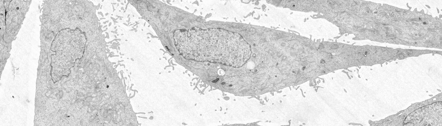
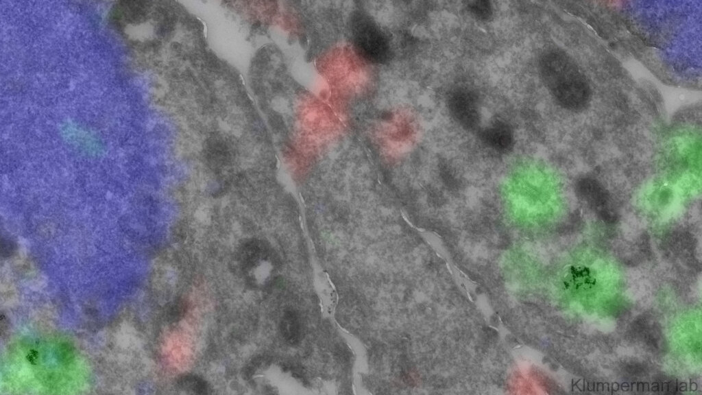
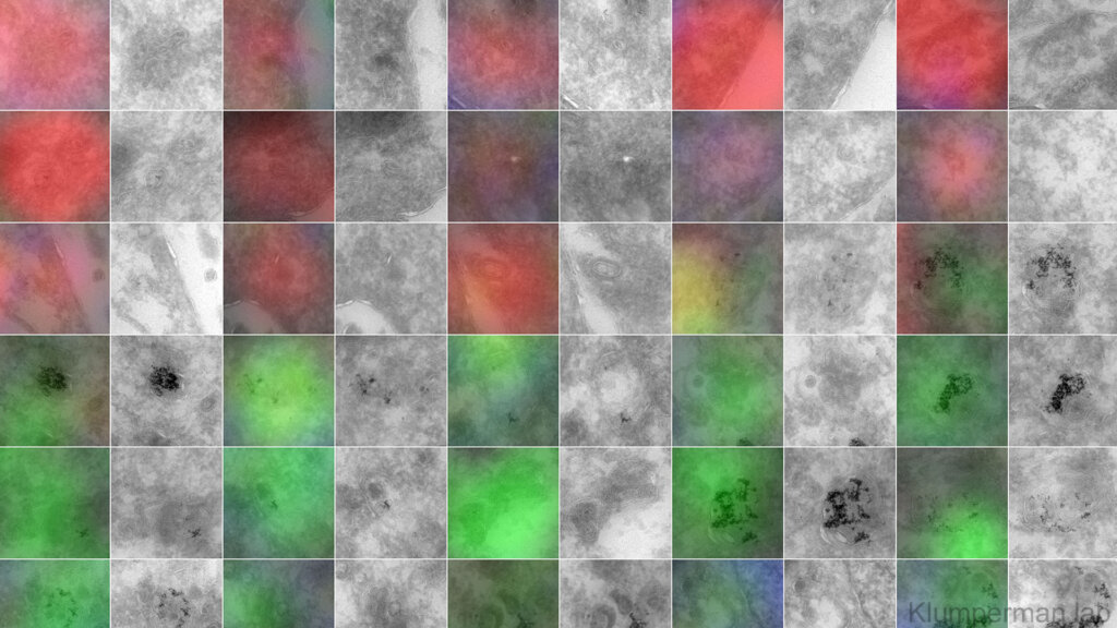
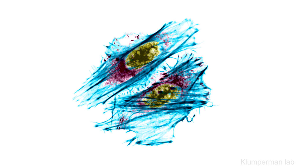
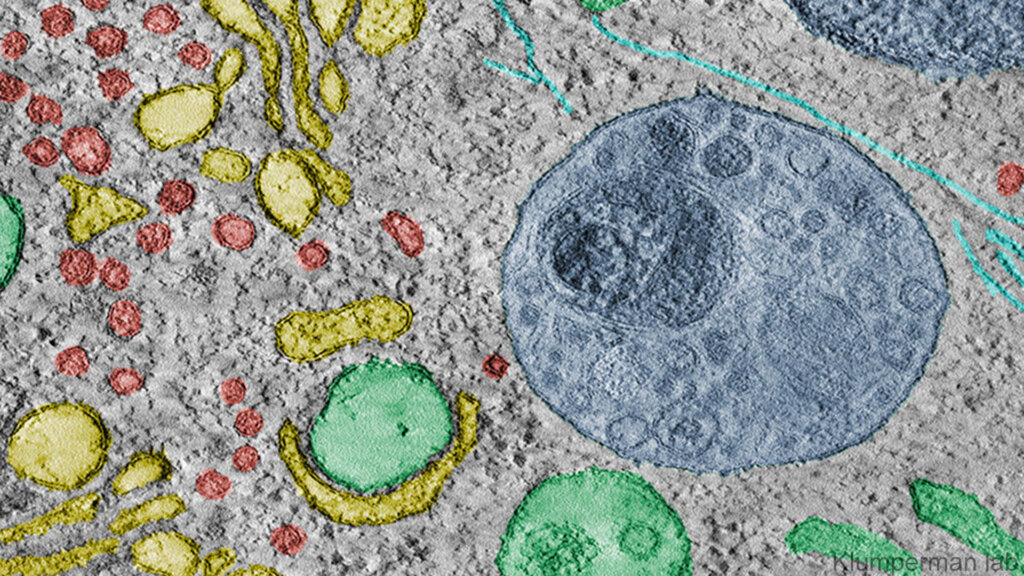
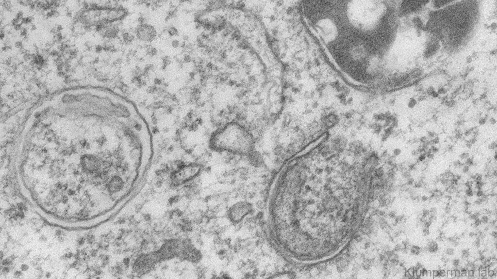
Cell Microscopy Core (CMC), is an expertise center for microscopy at the subcellular, cellular and tissue level. CMC is specialized in, and internationally renowned for immuno-electron microscopy (immunogold labeling of Tokuyasu cryosections) and correlative light-electron microscopy (CLEM). These methods uniquely allow the study of molecules at the nanoscale, within the ultrastructural context of the cell.
CMC is ratified as the CLEM node within the national Roadmap projects Netherlands Electron Microscopy Infrastructure (NEMI) and NL-BioImaging, as well as the European ESFRI project EuroBioImaging.
The infrastructure of CMC comprises equipment for EM sample preparation of biological specimens, fluorescent microscopy, live-cell imaging, transmission EM, FIB-SEM and CLEM. Together this covers a full range of microscopy methods for integrated studies at the subcellular, cellular and tissue level.
Expert CMC staff provide microscopy training, assistance with experimental design and interpretation, microscopy and imaging analysis courses, and full project service. This effectively allows users to address fundamental and applied research questions that require molecular and ultrastructural information.
Let’s connect!
Facility managerCorlinda ten Brinkcellmicroscopy@umcutrecht.nl088 - 57756548 Visiting address:Heidelberglaan 1003584CX Utrecht
Facility features:
- Conventional electron microscopy (EM)
- Immuno-electron microscopy (immuno-EM): the cryo-immunogold technique
- Electron tomography
- Scanning Electron Microscopy (SEM)
- Volume Electron Microscopy (VolumeEM)
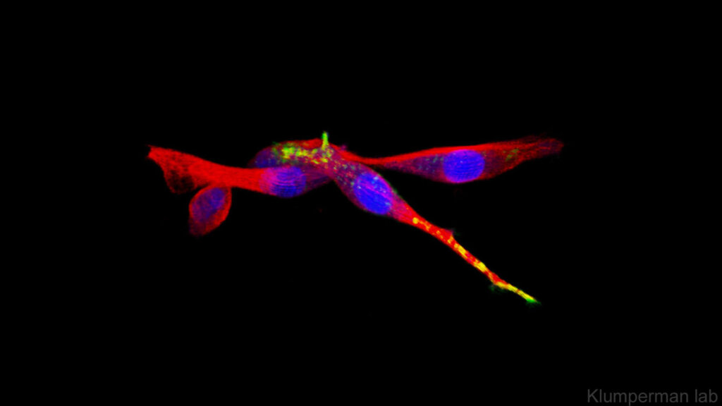
- Light microscopy, widefield, confocal, FRAP, FRET, Tirf
- Live-cell Imaging
- Live-cell Correlative Light- and Electron Microscopy (CLEM)
- Section CLEM
- High pressure freezing (HPF)