Princess Máxima Imaging Center
The Princess Máxima Imaging Center offers an advanced imaging infrastructure, expert training, and dedicated experimental support. Our mission is to drive research forward by providing cutting-edge imaging technologies and equipment, enabling researchers to capture the dynamic spatial organization of biological processes using state-of-the-art imaging capabilities.
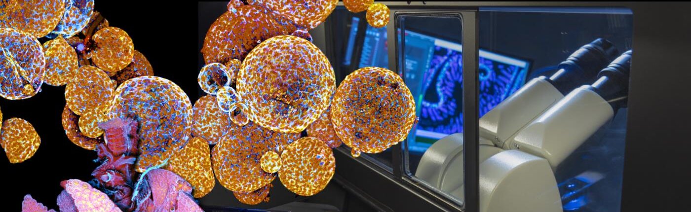
The Princess Máxima Imaging Center plays a crucial role in pediatric cancer research by providing state-of-the-art imaging technology and expertise. Microscopy plays a vital role in visualizing the morphology, behavior, and activity of cells over time, enabling a deeper understanding of spatial and temporal dynamics in complex biological systems. By investing in microscopy, researchers gain a unique perspective on the interactions and processes within 3D organoids. This facilitates the study of human biology, drug screening, and examination of cancer and environmental cell interactions, particularly in the context of immunotherapies.
Expert knowledge and support accelerate research through guidance in experimental design, image processing, and quantification. Collaboration and knowledge exchange through user trainings and workshops foster innovation and expedite discoveries. Outcomes include a better understanding of patient heterogeneity, biomarker identification, and insights into therapy resistance mechanisms.
The center empowers researchers with cutting-edge imaging technologies, expert support, and a collaborative environment, enhancing studies, improving outcomes, and advancing therapeutic strategies for children with cancer.
Equipment overview:
- M80 dissection microscope
- M205 FA automatized fluorescence stereomicroscope
- DM6 upright fluorescence microscope
- DMi8 widefield fluorescence microscope, suitable for live imaging
- DMi8 THUNDER widefield fluorescence microscope, suitable for live imaging
- SP8 confocal microscope with 8Khz resonant scanner, suitable for live imaging
- STELLARIS confocal microscope with 8Khz resonant scanner and WLL, suitable for live imaging
- LSM880 dual multiphoton/confocal platform equipped with AiryScan suitable for live imaging
- Nikon Ti-2 Eclipse spinning disk confocal, suitable for live imaging and autonomous microscopy
- STELLARIS FALCON confocal microscope with 8Khz resonant scanner and WLL, FLIM lifetime imaging, suitable for live imaging
- Zeiss LSM980 confocal microscope, suitable for live imaging and autonomous microscopy
- Revvity Opera Phenix Plus spinning disk confocal with 4 cameras for high-content screening, suitable for live imaging
- Zeiss Axioscan 7 slide scanner, suitable for brightfield and fluorescence
- Ramona Optics MCAM Vireo multicamera array microscope, suitable for brightfield or fluorescence-based high-content screening
- High-end image analysis workstations
The Princess Máxima Imaging Center supports the Center’s research groups and researchers via project collaborations. For external parties interested in accessing the facility, they are encouraged to reach out and contact the center for further information and potential collaboration opportunities
Let’s connect!
Facility managerRavian van Ineveldimaging@prinsesmaximacentrum.nl Visiting address:Prinses Máxima Center // Heidelberglaan 253584 CS Utrecht
Facility features: